Journal of Proteome Research

Viewing posts tagged Journal of Proteome Research

Li lab project uses DiLeu isobaric labeling to monitor neurotoxicity in chemotherapy in children

A new project by the Lingjun Li lab explores the use of the 4-plex N,N-dimethyl leucine (DiLeu) isobaric labeling strategy for protein identification and quantification in cerebrospinal fluid in children undergoing chemotherapy. The results from this and future studies will provide a means to monitor neurotoxicity and develop strategies to prevent central nervous system injury in response to chemotherapy in children.

The publication is Isobaric labeling strategy utilizing 4-plex N,N-dimethyl leucine (DiLeu) tags reveals proteomic changes induced by chemotherapy in cerebrospinal fluid of children with B-cell acute lymphoblastic leukemia from the Journal of Proteome Research.

Li lab collaboration explores noninvasive markers in prostate disease diagnosis

A recent collaboration between the labs of Lingjun Li and William Ricke explores the relationship between prostatic hyperplasia and related lower urinary tract symptoms in aging males and how noninvasive markers could be helpful in disease diagnosis. This proteomics study used a mouse model of hormone-induced urinary dysfunction to gain insight into the disease and supports the concept of noninvasive urinary biomarkers being a successful route for prostate disease diagnostics.

Thomas S, Hao L, DeLaney K, McLean D, Steinke L, Marker PC, Vezina CM, Li L, Ricke WA. Spatiotemporal proteomics reveals the molecular consequences of hormone treatment in a mouse model of lower urinary tract dysfunction. Journal of Proteome Research. 2020, 19(4):1375-1382.

Quantification of the Human Pancreatic ECM

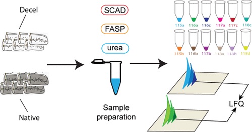
Ma et al (2019) recently published a paper on the quantification of human pancreatic extracellular matrix proteins in the Journal of Proteome Research.

In this study, researchers characterized the composition of the human pancreatic extracellular matrix (ECM) before and after decellularization. To find the relative quantification of ECM proteins, they used isobaric dimethylated leucine (DiLeu) labeling.

It was important for researchers to look at the ECM of the pancreatic microenvironment as it is essential to pancreatic function– it regulates β cell proliferation, differentiation, and insulin secretion.

As a result of decellularization, and through quantitative proteomic analysis, most cellular proteins were removed while matrisome proteins remained. This process generated a large data set of matrisome proteins from a single tissue type. 

Researchers then quantified the distinct expression of ECM proteins, comparing adult and fetal pancreas ECM. This revealed a correlation between matrix composition and postnatal β cell maturation.

Overall, the results of this study sheds light on the prospect of bioengineering a pancreas. Additionally, the study demonstrates the roles that matrisome proteins have in postnatal β cell maturation.

Graphical abstract for Ma et al (2019), depicting native and decelled pancreatic extracellular matrix proteins, sample preparation technique, and LFQ

CZE and Mass Spectrometry Leads to Considerable Phosphopeptide Identification

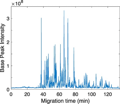
Zhang et al demonstrated the importance and utility of Single-Shot Capillary Zone Electrophoresis in the Journal of Proteome Research.

Capillary zone electrophoresis (CZE) is a practical tool in exploring and interpreting post-translational modifications in proteins. To examine the usefulness of single-shot CZE with mass spectrometry through the analysis of phosphoproteomics, researchers used CZE separations with the Orbitrap Fusion Lumos Tribrid platform, and used a linear-polyacrylamide-coated capillary with low electroosmotic flow for separation.

Researchers found that larger injection volumes led to broader peaks and less phosphopeptide identifications. Additionally, in this single-shot phosphoproteome analysis, researchers found 4405 phosphopeptides out of an original 220 ng enriched phosphopeptides from a mouse brain.

Data for this study is available in the ProteomeXchange with identified PXD012888.

Graphical abstract for Zhang et al. This graph shows how Base Peak Intensity compares to Migration Time (in minutes). It appears that base peak intensity is higher during the 38 minute mark, the 63 minute mark, and the 65 minute mark.